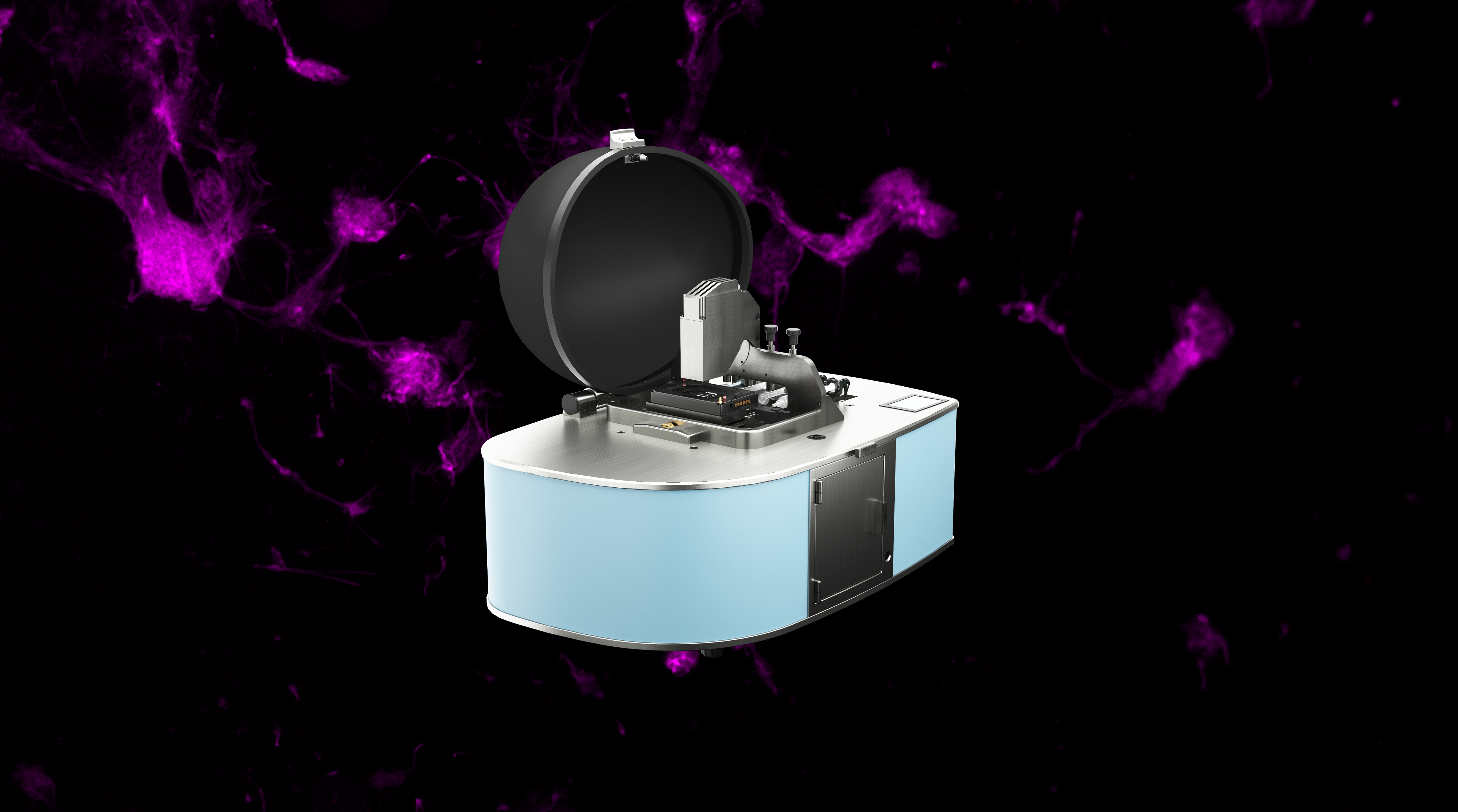
Discover the D-Tails product
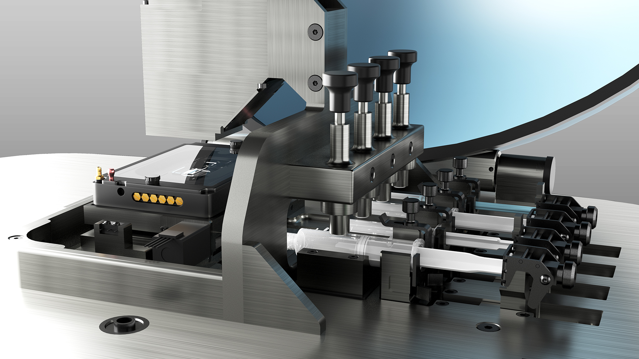
A cutting-edge integrated system
Our latest innovation in the field of microscopy and microfluidics
A groundbreaking platform for a new era in wide-field image acquisition
iFLUOR: Revolutionary Imaging and Microfluidics
iFLUOR test is an integrated system that brings together advanced wide-field fluorescent microscopy with microfluidic technology, offering researchers a powerful tool for high-throughput, long-term imaging of live cells and small organisms such as C. elegans. It addresses critical challenges in cancer diagnostics, neurological research, and other fields where both detailed imaging and large population analysis are required.
Its ultrawide field of view (10x10 mm) with a pixel resolution of 2.2 microns, paired with advanced fluidic control, allows for uninterrupted, real-time observation of cellular responses under various conditions. The seamless integration of super-resolution imaging and microfluidic channels enables precise control over experiments without perturbing samples, ensuring more accurate results.
Key Features of iFLUOR
Discover the features that redefine what's possible in fluorescence imaging
Ultrawide Field of View
10x10 mm with a remarkable 2.2 microns pixel resolution

Capture large populations of cells or organisms with a wide 10x10 mm imaging area at high resolution, ideal for statistical analysis and large-scale experiments.
Versatile Imaging
Capabilities for transmission imaging and single-color fluorescence
Supports both transmission imaging and single-color fluorescence, enabling researchers to view the same sample in multiple ways without moving the sample.
Advanced Microfluidics
Up to 4 channels with integrated microfluidic pumps
Up to 4 integrated microfluidic channels with programmable fluidic delivery for real-time control of the environment, perfect for dynamic, long-term cell experiments.
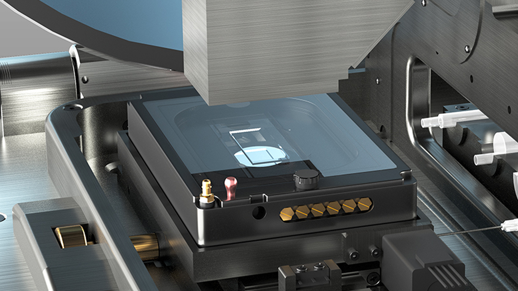
Enhanced Adaptability
Includes a micro incubator adapter
Compatible with customizable chip designs for specific research needs, such as barrier-controlled environments for small organisms like C. elegans.
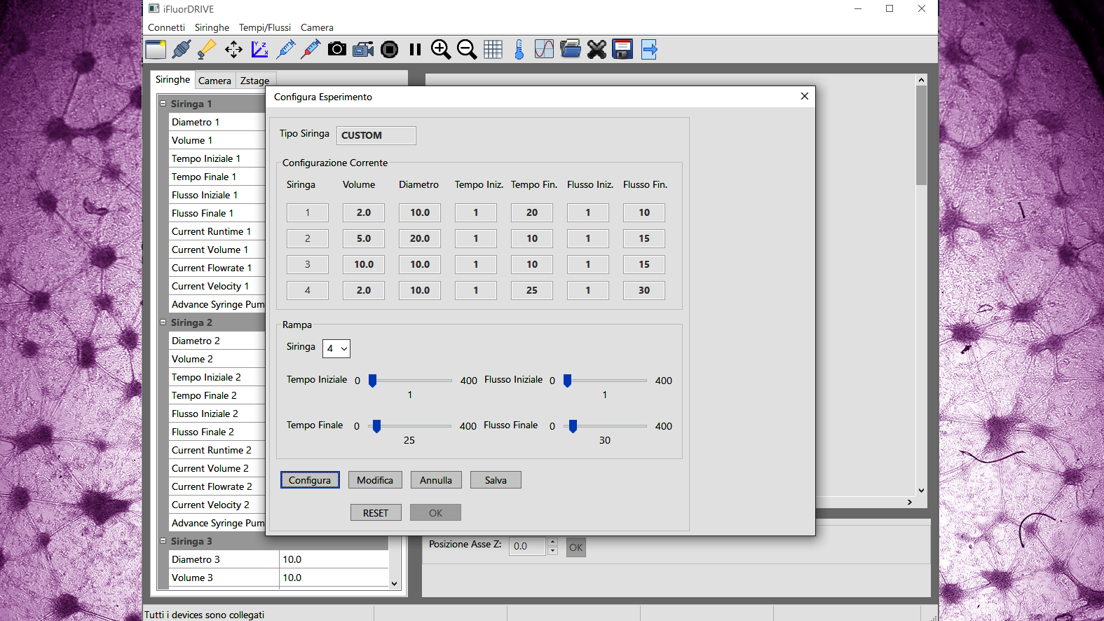
All-in-one Software
Timelapse design, timed and/or ramp fluidic delivery, acquisition controls, super resolution image acquisition and data analysis

The all-in-one software allows for timelapse design, automated image acquisition, data analysis, and control over microfluidic delivery, simplifying complex experiments.
Super-Resolution Imaging
Enhances imaging with sub-micron accuracy over a wide field
Achieve a pixel resolution of 0.6 microns with the iFLUOR Pro version, using proprietary micro-scanning technology for unmatched detail across the entire imaging field.
Upgradeable Options:
Advanced features for enhanced performance and flexibility
Extended Scanning
X scanning unit for multiwell acquisition up to 40 mm
The extended scanning option allows seamless multiwell acquisition, providing flexibility for high-throughput experiments. This upgrade is ideal for laboratories needing precise scanning over larger areas.
Enhanced Incubation
Fully integrated micro incubator
Our fully integrated micro incubator ensures that your samples are maintained in optimal conditions for long-term experiments, with precise control over temperature and environment.
Dual-Color Fluorescence
Expands imaging capabilities
The dual-color fluorescence option expands your imaging capabilities, enabling simultaneous detection of multiple markers. This upgrade is crucial for complex multi-channel experiments.
Professional Version
Features proprietary micro-scanning and AI software achieving 0.6 microns pixel resolution across the entire 10x10 mm field
Unlock the full potential of iFLUOR with the Professional Version, featuring AI-driven micro-scanning technology that enhances pixel resolution to an impressive 0.6 microns across the entire field.
The standout feature of the iFLUOR is its seamless integration of the microfluidics system with the 10x10 mm field of view and high resolution. This allows for uninterrupted imaging of cell populations without the need for sample movement, thereby reducing the risk of perturbations and bubble formation. Additionally, an integrated leak detector provides immediate alerts, even remotely, ensuring the reliable performance of the microfluidics circuit.
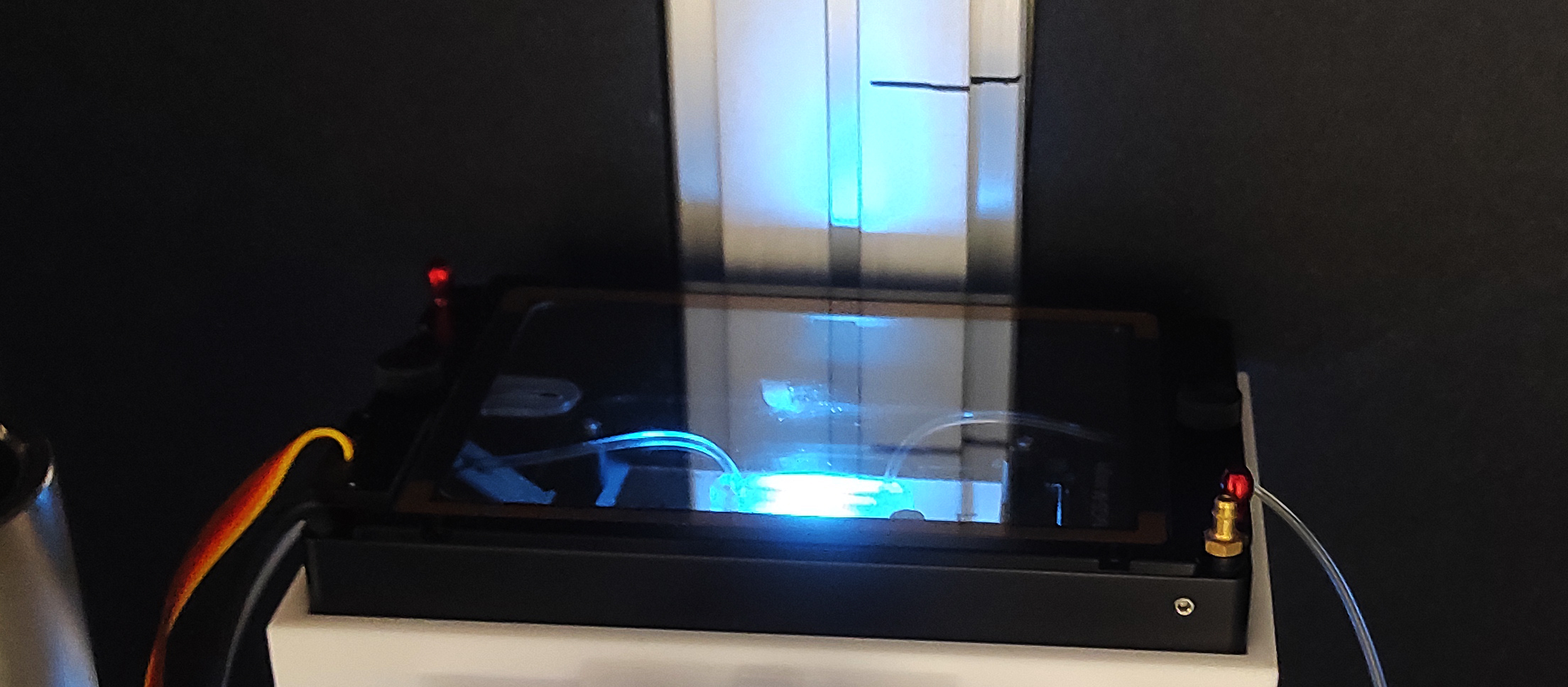
The iFLUOR’s design includes an integrated cover, enabling image acquisition even in lit environments.
Applications of iFLUOR
iFLUOR offers numerous possibilities for research across various fields due to its versatile imaging capabilities and advanced microfluidics system
Neural rosettes:
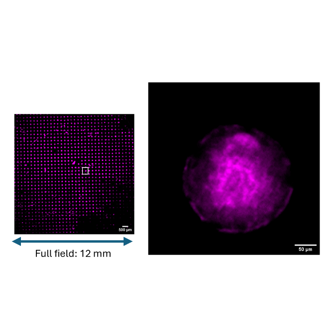
Ultra-wide field acquisition of hundreds of human iPSC-derived neural rosettes on micropatterned support.
At D-tails we are working to make ultra-wide field imaging with enhanced resolution accessible to all researchers and to advance their scientific potential and breakthroughs to the next level. Our new product, the iFluor system, is based on a relay lens (Edmund Optics #1937) able to acquire multicellular structures up to a 10x10 mm field of view, hosting different sample holders. A set of standout features make iFluor a versatile and adaptable system, including capabilities for transmission imaging and single-color fluorescence, possibility of equipping up to 4 channels with integrated microfluidic pumps, presence of a micro incubator adapter, availability of all-in-one software that allows timelapse design, timed and/or ramp fluidic delivery, acquisition controls, super resolution image acquisition and data analysis. In this Application Note, we demonstrate that iFluor system is able to acquire a 10x10mm surface containing 900 experimental replicates, at 2.2 microns pixel resolution and 1x magnification. The “SR” version is also able to super-resolve a digitally magnified single replicate up to 0.6 microns pixel, thanks to a custom algorithm for super-resolution...
Look at the image...
Representative images of full-field acquisition (left) of neural rosettes immunostained with Atto 532-Phalloidin (Sigma Aldrich). Scale bar 500 µm (n= 900 rosettes). Zoomed-in and super-resolved image (right) shows a single neural rosette. Scale bar 50 µm.
Organoids:
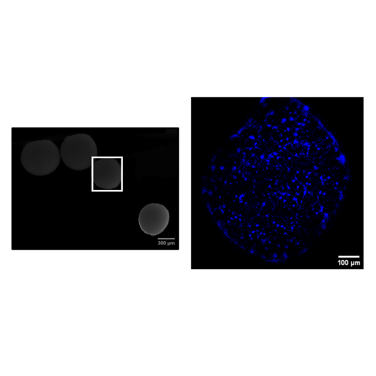
iFLUOR's advanced microfluidics system facilitates the growth, maintenance, and imaging of organoids under controlled conditions
iFLUOR’s microfluidics technology enables precise control over the growth environment, making it an essential tool for studying complex 3D cell structures like organoids. Researchers can simulate and monitor development in real-time, providing critical insights into organ formation and disease modeling.
Look at the image...
Acquisition of cortical organoids with anti-Pax6 antibody (Santa Cruz Biotechnology) (left). Zoomed-in image (right) shows a single cortical organoid. Scale bar 100 µm.
Super resolution:
The iFLUOR’s pro super-resolution algorithm enhances minute details in the ultra-wide field, resulting in significantly improved image quality
With iFLUOR’s super-resolution algorithm, researchers can achieve incredibly detailed imaging, even at sub-micron levels. This feature is particularly beneficial for visualizing complex structures in tissues or cellular components that require high-definition imaging for accurate analysis.
Look at the image...
Super-resolution imaging of retinal neurons immunostained with Atto 532-Phalloidin (Sigma Aldrich). The images show the same field: simple acquisition (left) and super-resolved images (right). Scale bar: 100 µm.

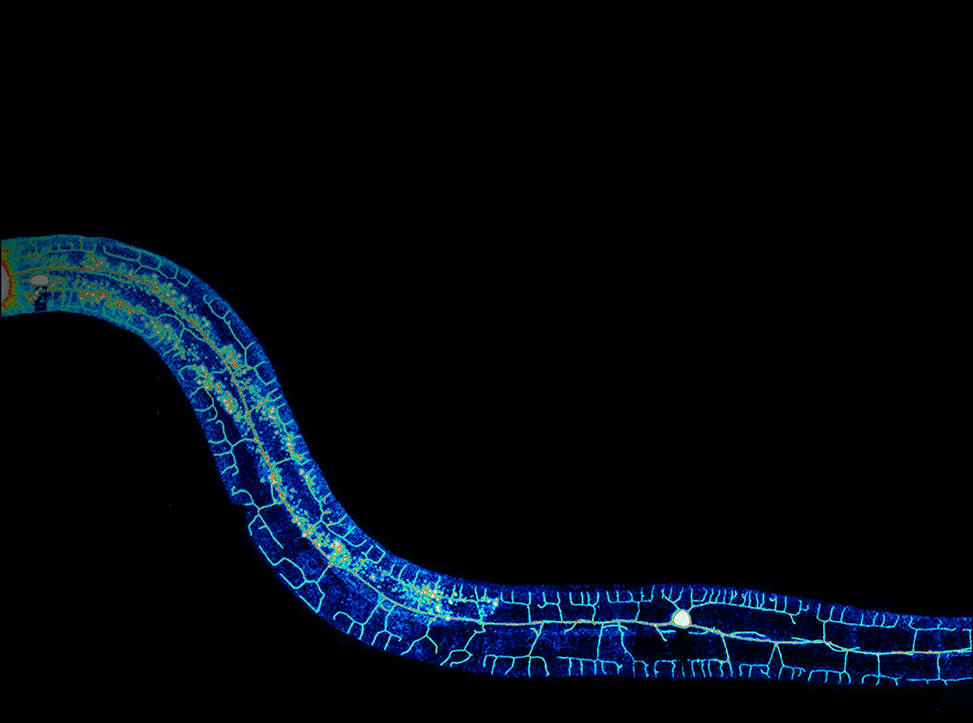
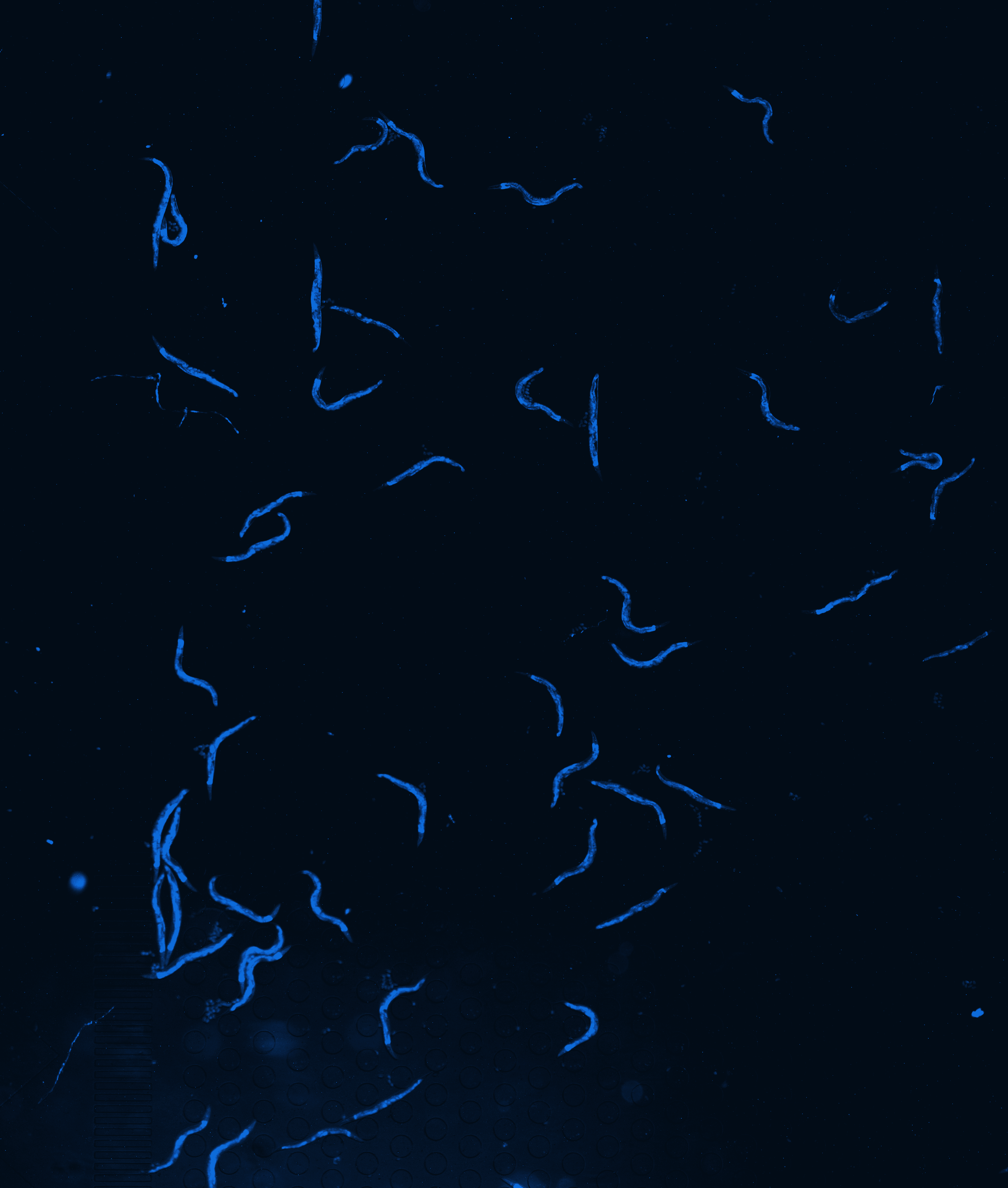
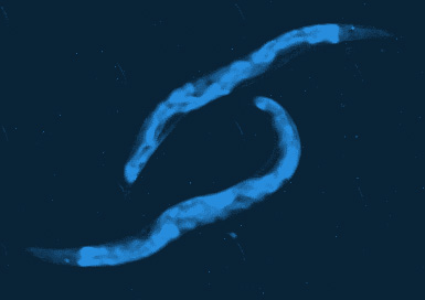
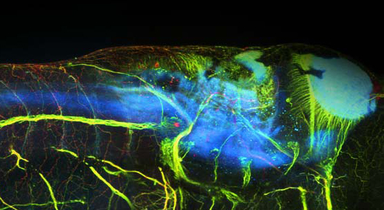
C. elegans:
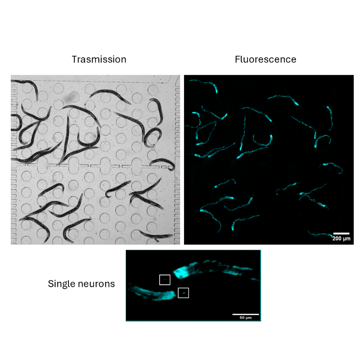
Ultra-wide field acquisition of a population of C. elegans in a microfluidic chip.
At D-tails, we developed an innovative acquisition system that boasts ultra-wide field of view coupled with high-resolution imaging capabilities, that enable researchers worldwide to acquire images across diverse applications in a groundbreaking way. Our new product, the iFLUOR system, is based on a high-quality relay lens and a cutting-edge CMOS sensor able to acquire images with a field of view of 10 x 10 mm2, paired with an illumination system that includes two customizable channels (transmission and fluorescence). In addition to that, our device integrates a syringe pump system with up to four syringe pumps equipped with temperature and leakage controls, for managing microfluidic experiments, maintaining cell cultures, or preserving sample integrity during in vivo experiments. For a better experimental control, iFLUOR is also compatible with Okolab incubators. Finally, its dedicated software allows for timelapse design for up to 3 days, fluidic delivery with customizable dynamics, acquisition controls, super resolution image acquisition and data analysis. In this Application Note, we use the iFLUOR system to acquire full-field images of a population of C. elegans loaded in a microfluidic chip, using the transmission and fluorescence channel at 2.2 microns pixel resolution and 1x magnification.
Look at the image...
Transmission channel (left) and fluorescence channel (right) images of C. elegans with calcium indicator expression in a microfluidic chip. Scale bar 200 µm. Zoomed-in image shows single neurons with calcium indicator fluorescence in the head of C. elegans. Scale bar 50 µm.
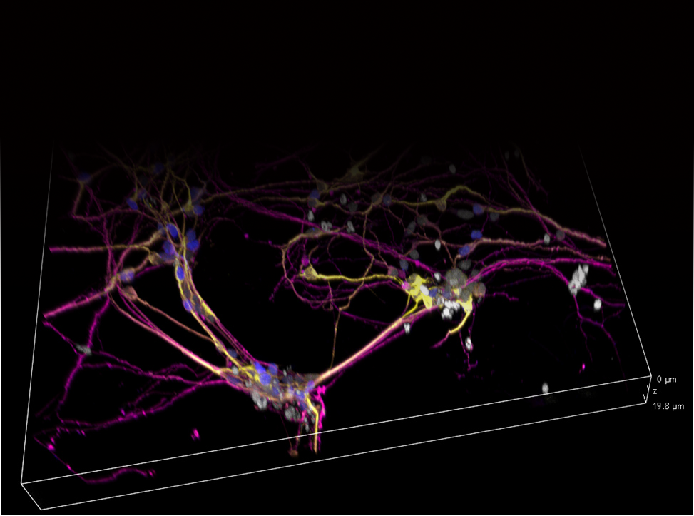
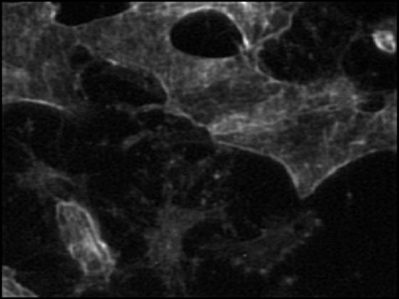
Activity dynamics in neural cultures:
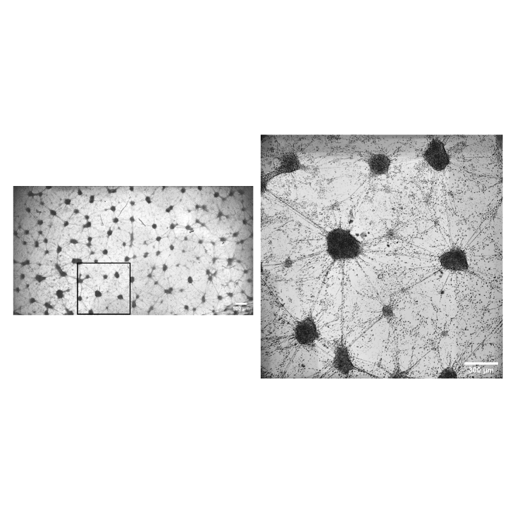
iFLUOR can monitor neuronal activity over large areas and extended periods
iFLUOR’s wide-field imaging capabilities make it ideal for monitoring activity in large populations of neurons over extended periods. Researchers can observe dynamic changes in neural activity in real-time, providing critical insights into brain function and disease.
Look at the image...
Full-width acquisition of retinal cultures during neural activity monitoring with Fluo4am (left). Scale bar 500 µm. Zoomed-in image of neural network (right).
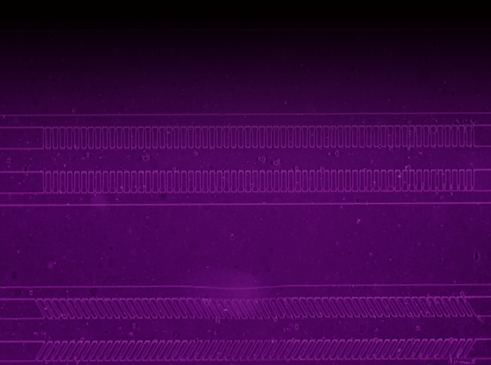
Cell growth dynamics:
iFLUOR enables long-term time-lapse imaging of cellular processes such as growth and division over several days, maintaining constant inflow of growth medium
iFLUOR supports long-term, time-lapse imaging of cell growth and division, enabling researchers to track cellular dynamics over several days. Its real-time fluid control ensures consistent experimental conditions, making it perfect for high-precision studies.
Look at the image...
Jurkat cell growth experiment in microfluidic chip. Scale bar 1 Mm.

Applications
Cancer Diagnostics
With its ability to track cell populations and individual cell responses, iFLUOR is ideal for cancer research. Its ultra-sensitive fluorescence detection can identify changes in biomarkers at early stages, making it invaluable for early diagnosis of cancers like breast, prostate, and pancreatic cancer.
Neuroscience Research
Utilizing the integrated microfluidics and real-time imaging capabilities, iFLUOR facilitates studies on neural activity. It has been instrumental in research involving C. elegans, with real-time calcium imaging and behavioral monitoring.
Organ-on-Chip Models
iFLUOR's ability to handle complex organoids and organ-on-chip models makes it a powerful tool for studying cell growth, organ development, and personalized medicine applications.
Real-World Application: Neural Activity Monitoring
iFLUOR excels in long-term, high-resolution studies of neural dynamics. For instance, in one application, it was used to monitor the activity of genetically modified C. elegans populations. These organisms were exposed to chemical stimuli, and their neuronal activity was tracked using fluorescence imaging, allowing researchers to observe how individual neurons responded to stimuli without disturbing the sample.
The integration of wide-field imaging with precise microfluidic control allowed the researchers to manage large populations of nematodes while also capturing fine details of individual neuronal responses. This made iFLUOR a crucial tool in the study of how neural networks react to different environmental stimuli, such as drugs or disease markers.
Transform Your Research with iFLUOR
Ready to upgrade your lab’s capabilities? Contact us for a demo or more information on iFLUOR.